
Images from Spring 2017 National ACS Meeting (Abstracts follow photos)


Abstracts from our group for the 253rd San Francisco ACS Meeting and Exposition: April 2, 2017 – April 6, 2017
The members in the Crans group gave serveral presentations at the 253rd ACS Meeting in San Francisco these presentations ranged from poster presentations to oral presentations. These presentations were in multiple divisions ranging from inorganic to biological chemistry. The abstracts of each presentation can be found below.
Division: ANYL – Analytical Division
Poster Number and Title: 227 – Photolysis of a Zn(II)-nitriliotriacetate cage in AOT/isooctane reverse micelles
Authors: Cheryle Beuning Twinkle Paryani, Noah Barkley, Prem Basa, Richard Cole, Nancy Levinger, Shawn Burdette, Debbie Crans
Abstract: We have designed a photolysis system in reverse micelles to study fast time-scale metal chelation by peptides within a membrane model system with the specificity to release metal ions at specific time by irradiation alone without having to manually add other solutions. Zn(II) ion release by photo-induced decarboxylation reactions of Zn(II)-nitriliotriacetate decarboxylation cage (Zn(II)-NTAdeCage) photo-complexes within confined spaces are governed by specific location of the compound and the charge compatibility of complex with the reverse micelle interface. Zn(II)-NTAdeCage has been shown in buffered aqueous solution (pH 7.5, 40 mM HEPES/100 mM KCl) to photolytically release Zn(II) with irradiation at 365 nm. The intact cage has a high affinity for Zn(II) and upon irradiation creates two photoproducts (iminodiacetic acid, m-nitrobenzaldehyde) which have little affinity for Zn(II). The current work shows successful photo-induced decarboxylation of the Zn(II)-NTAdeCage complex within AOT/isooctane reverse micelles (pH 7.5, 40 mM HEPES, 0.02 M AOT, w0 30, λirr355 nm). We observe the release of Zn(II) ions from Zn(II)-NTAdeCage complex by a decrease in the absorbance at 270 nm (Zn(II)-NTAdeCage) as the complex is degraded and the subsequent retrieval of Zn(II) by Zincon monosodium salt with peak growth at 620 nm of the Zn(II)-Zincon complex and peak loss at 470 nm (aqueous) or 540 nm (reverse micelle) of the free Zincon ligand in both aqueous buffered and reverse micelle systems.
Division: INOR – Inorganic Division-Oral Presentation
Presentation Number and Title: 1323 – Cu(II) coordination differences in the amyloid-beta protein binding models GHK and DAHK result in varied conditional inter-peptidic metal exchange rate constants
Authors: Cheryle Beuning, Béatrice Mestre, Christelle Hureau, Debbie Crans
Abstract: Inter-peptidic metal exchange is becoming increasingly apparent in neurological disorders like Alzheimer’s disease as amyloid-beta and other intrinsically disordered peptides sequester metals in the brain even from other metalloproteins. The short peptides GHK and DAHK and their fluorescent counterparts GHW and DAHW are known Cu(II) amyloid-beta binding models. These peptides are all strong Cu(II) chelators that have conditional (pH 7.4, 0.1 M HEPES) binding constants, Ka, of GHK (3N+1O binding), DAHK (4N binding) and amyloid-beta at 1.4 x 10¹³, 3.9 x 10¹³M-1 and at 2.4 x 10⁹ M-1, respectively. Using sensitive fluorescence spectroscopy and taking advantage of proximity fluorescence quenching of tryptophan by Cu(II), we have monitored the exchange from Cu(II)-GHW to GHK and DAHK rather efficiently. The conditional (pH 7.4, 0.1M HEPES) second order rate constants of two exchanges from Cu(II)-GHW to GHK and DAHK were determined to be 1.4 ± 0.2 x 10² and 4.7 ± 1.9 x 101 M-1s-1, respectively. We propose an intermediate ternary GHW-Cu(II)-GHK/DAHK complex likely facilitates the exchange as the hydrated coordination spot in the Cu(II)-GHW complex can undergo a ligand-exchange with the incoming peptide’s N terminus or imidazole N’s. Changes in the superhyperfine coupling lines in the EPR spectra in experimental conditions (1:2 vs 1:1 Cu:GHK) do support the formation of the ternary complex where in 1:2 Cu:GHK conditions a 4th N-donor ligand binds when the second peptide is added to Cu(II)-GHK instead of an O-donor as seen in 1:1 Cu:GHK conditions. These studies show that metal coordination plays an important role in metal exchange kinetics and we plan to extend this method development to study inter-peptidic metal exchange of amyloid-beta with itself and other biologically relevant peptides.
Division: BIOL – Biological Chemistry Division
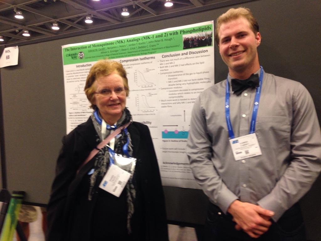
Poster Number and Title: 68 – Interaction of Menaquinone (MK) analogs (MK-1 and MK-2) with phospholipids
Authors: Gabriel Cardiff, Benjamin Peters, Jordan Koehn, John Peter Hough, Allison Groninger, Dean Crick, Debbie Crans, Fabio Levi Fontes
Abstract: Tuberculosis is a worldwide epidemic that is caused by Mycobacterium tuberculosis. With the rise of drug resistant Mycobacterium tuberculosis, it has become increasingly important for a better understanding of the interaction between substrate analogs of virulance factors with membranes. A potential novel virulence factor, MenJ, has been shown to selectively reduce the second isoprene unit of the electron carrier, menaquinone (MK-9), within the electron transport chain. As MK-9 is membrane bound, it is of interest to determine the membrane interactions of the napthaquinone of MK-9. To probe the interactions of the napthaquinone of MK-9 with membranes, we explored the interactions of MK-9 analogs with only one or two isoprene units (MK-1 and MK-2, respectively) with phospholipid monolayers. Through this work, we have found differences in interactions of MK-1 and MK-2 with different lipids including dipalmitoylphosphatidylcholine (DPPC) and dipalmitoylphosphatidylethanolamine (DPPE).
Division: INOR
Presentation Number and Title: 1210 – Controlling reactivity of small molecules through confinement
Authors: Michael Johnson, Debbie Crans
Abstract: The reactivity and speciation of small molecules in confined media, such as microporous materials, cellular substructures or membrane surfaces, have been assumed to be similar to those found in bulk solvents. Part of the reason for this assumption is the difficulty to study molecules and their reactions in confinement. Over the past several years we have used reverse micelles (RMs) to study reactions and speciation within carefully confined media. These models provide a well defined and controlled series of spherical nanodroplets of solvent water homogeneously dispersed within organic solvents. Sizes, numbers of waters, surface charges, etc. are known and these parameters can be easily altered to provide a series of confined structures in which to study molecular reactivity and speciation. For example, we have observed significant enhancement of electron transfer rates between ascorbic acid and iron(III) centers and have worked to define the changes in reaction conditions, molecular speciation and properties as well as location within the RM structures to understand the observed reactivity changes. The results of our work are presented.
Dr. Crans also organized and presided over the INOR: Celebrating 60 years of inorganic chemistry Symposium that ran from April 2nd to the 5th.
Division: INOR and in Sci-Mix poster session
Poster Number and Title: 226 – Selenium speciation in the Fountain Creek water and effects on fish species diversity
Authors: Jim Carsella, Sandra Bonetti, Debbie Crans, Scott Herrmann, Delwayne Nimmo
Abstract: Speciation is important to the environmental effects of selenium, isoelectronic to sulfur, and consideration of the particular speciation under environmental conditions allows for identification of beneficial and more toxic forms. The most common forms of Se in the aquatic environment are the oxidized forms selenate (Se(VI)) and selenite (Se(IV)). These forms of Se enter the water as a result of oxidation and weathering of substrate minerals. Recent evidence indicates that the presence of salts and nitrates can change the forms of Se present in natural waters in a way that makes current Pourbaix diagrams inaccurate when working with low Se concentrations present in rivers1. This inaccuracy requires Se species to be measured by LC-ICPMS and may be used to determine the toxicity of these forms in natural water ecosystems1. Se is a dietary requirement for all animals and humans and can be toxic if present in high concentrations. The EPA has listed the chronic exposure limits for fish tissue as 15.8 mg Se/Kg for egg/ovary, 8.0 mg Se/Kg whole body. The new chronic water exposure limit is under agency review and may be lowered to 1.2 mg Se/L. The Fountain Creek Watershed has segments of Fountain Creek where Se water levels exceed the current EPA limits of 5 mg/L. The source of this Se is the Se rich Pierre Shale. According to Pourbaix diagrams, the Se species present should be hydrogen selenite. However our results indicate that the Se present is the less toxic selenate. The high levels of Se present in the creek and the native bryophytes would predict negative trends in the health and the diversity of fish present at sites with high Se. The results of our analysis show that fish diversity is unaffected by the Se levels in the Fountain Creek. This result indicates that other factors may be involved in the availability of Se to aquatic organisms in Fountain Creek.
Division: BIOL
Poster Number and Title: 214 – Interfacial interactions of glycine and short glycine peptides in confined spaces
Authors: Kaitlin Doucette, Prangthong Chaiyasit, Donn Calkins, Kayli Martinez, Mary Fisher, Debbie Crans, Anan Tongraar
Abstract: Understanding the interactions of small molecules at interfaces is critical for understanding uptake of metabolites and their availability at cellular uptake receptors. In this study, glycine and the short glycine peptides diglycine, triglycine, and tetraglycine are studied with regards to their interactions at the membrane mimetic interface of reverse micelles via 1H NMR and DLS measurements. It was found that with the exception of monomeric glycine, the peptides prefer to associate themselves with the interface of the reverse micelle. Monomeric glycine, however has been shown to locate itself with the N-terminus in the ordered interstitial water of the reverse micelle between the interface and the bulk water pool, with the C-terminus located in the bulk water pool.
Division: INOR and in Sci-Mix Poster session
Poster Number and Title: 968 – Vanadium-Dependent Haloperoxidase Enzymes: A Review of Mechanism, Structure-Functional Relationships, and Coordination Environments
Authors: Kaitlin Doucette, Craig Wallace, Craig McLauchlan, Debbie Crans
Abstract: Haloperoxidases are a class of enzymes that catalyze the reactions of halides with peroxides to form water and halogenated products. These products can be hydroxyhalides (XOH) or halogenated organic compounds (RX) but will react rapidly in biological systems. Primarily found in marine algae and bacteria or terrestrial fungi, haloperoxidases are divided into three classes: heme-dependent, non-heme-dependent, and vanadate-dependent. Using reported X-ray crystal structures of the vanadate-dependent haloperoxidases, the coordination environments, structure-function relationships, and enzyme halide specificity of the vanadium center are reviewed here. All but two of the coordination environments from the reported crystal structures of vanadium dependent haloperoxidases are trigonal bipyramidal with a tau value of greater than 0.7. Suggested mechanism of action is a nucleophilic attack of the peroxide to form an oxohalide, with the final step either halogenation of an organic compound or reaction with excess hydrogen peroxide to form water and the corresponding halide acid, which is consistent with proposed mechanisms.
Division: MEDI – Medicinal Chemistry Division
Poster Number and Title: 201 – Interactions of isoniazid with model membranes
Authors: Allison Groninger, Benjamin Peters, John Peter Hough, Fabio Levi Fontes, Gabriel Cardiff, Dean Crick, Debbie Crans
Abstract: Despite being a curable disease, tuberculosis (TB) is still one of the world’s most common diseases with an estimated one third of the population infected with latent TB. A current first line tuberculosis drug is isoniazid. The mechanism of the drug is known in that it couples with NAD+ to form a complex that can bind to the enoyl-AcpM substrate and the action of fatty acid synthesis. This inhibits synthesis of mycolic acids effectively inhibiting growth of mycobacterium tuberculosis. It also inhibits cytochrome P450 system and thus can be a source of free radicals. Though we know the function of the drug, the method of uptake into the cell is unknown. Since isoniazid is a small molecule it may be able to diffuse through the membrane but there is limited information about the interactions of isoniazid with membranes. To explore this membrane interaction, the interactions of isoniazid with reverse micelles and phospholipid Langmuir monolayers were studied at different pH values. Through this research, it was discovered that isoniazid interacts with various phospholipids differently and the exact placement of isoniazid at a surfactant interface was determined.
Division: MEDI
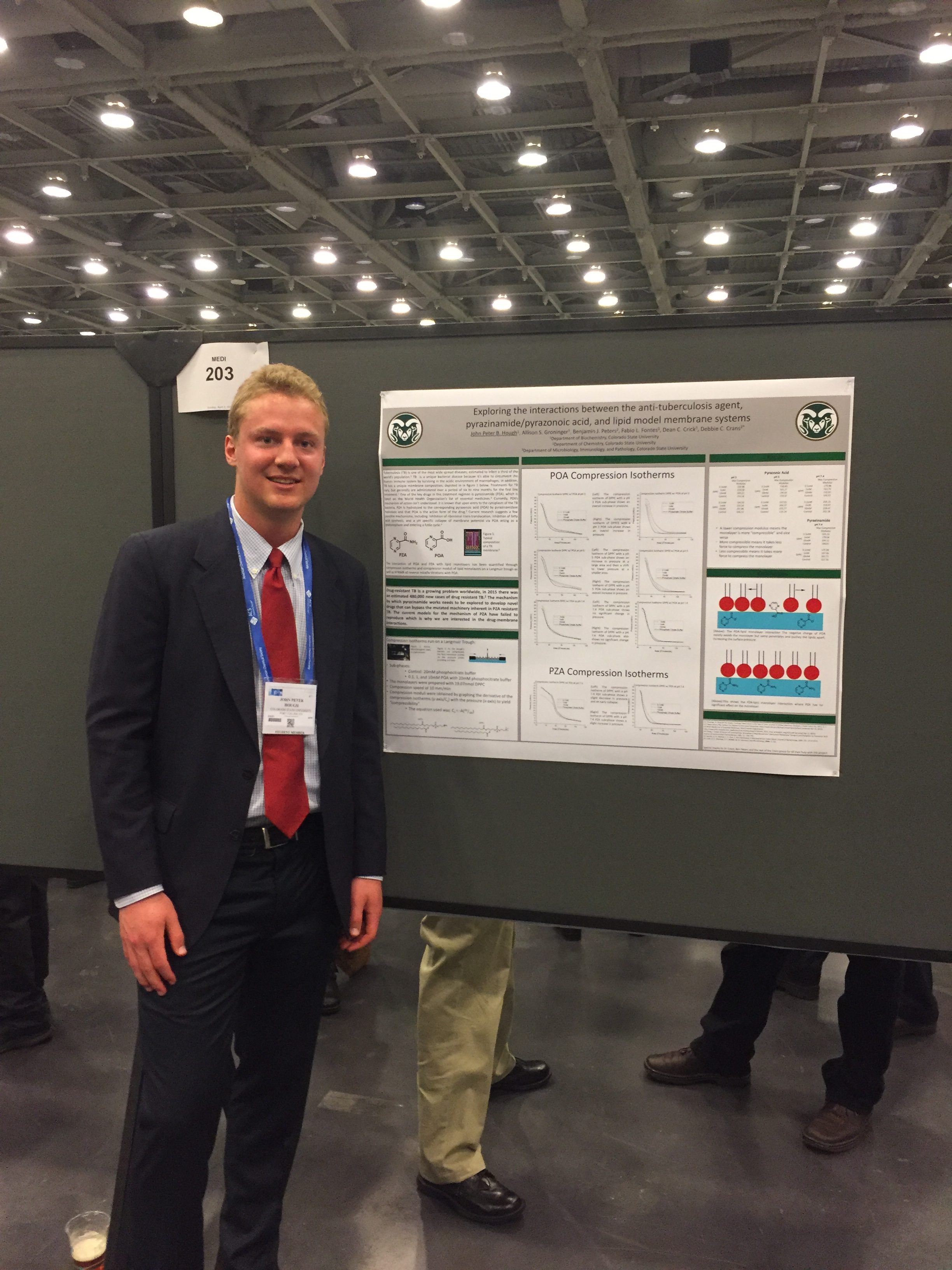
Poster Number and Title: 203 – Exploring the interactions between the anti-tuberculosis agent, pyrazinamide/pyrazonoic acid, and lipid model membrane systems
Authors: John Peter Hough, Allison Groninger, Benjamin Peters, Fabio Levi Fontes, Dean Crick, Debbie Crans
Abstract: Pyrazinamide (PZA) is a drug listed on the World Health Organization’s list of essential medicines due to its crucial role in treating tuberculosis. PZA is known to enter the cell and be hydrolyzed into pyrazonoic acid (POA). Once hydrolyzed, it has been shown to do two things: collapse the membrane potential, and inhibit ribosomal rescue. We are interested in the membrane interaction due to this collapsing of the membrane potential. To look at this, NMR studies of reverse micelle microemulsions were used to identify the location of PZA/POA in a model of a membrane leaflet. Interactions between POA/PZA and Langmuir monolayers, prepared from various phospholipids, were also measured using a Langmuir trough. These studies help characterize the drug-membrane interaction, which provides insight into PZA’s mechanism of disrupting membrane potential.
Division: INOR and in the Sci-Mix Poster Session
Poster Number and Title: 233 – Correlating redox potential with 51V NMR chemical shifts for vanadium (V) catecholates
Authors: Jordan Koehn, Pabitra Chatterjee, Cheryle Beuning, Andrew Waterhouse, Taylor Lucia, Tatyana Polenova, Debbie Crans
Abstract: Herein, we demonstrate the correlation of 51V NMR chemical shifts with redox potentials for a series of non-innocent vanadium(V) catecholate complexes. Several of the synthesized complexes shown here were characterized previously by both solid state and solution 51V NMR spectroscopy. Illustrated is an observed linear correlation between solution and solid state 51V NMR chemical shifts, which is consistent with the solution structure being similar to that observed in the solid state. Since the solid state 51V NMR chemical shift describes the electronic structure of the vanadium complexes, there exists the potential for the use of the 51V NMR chemical shift to characterize the properties of these complexes. The vanadium(V) catecholate complexes used in this study are, in fact, redox active and the chemical shifts fall outside the normal populated isotropic 51V NMR chemical shift range (-300 to -700 ppm), which is anticipated for complexes with non-innocent redox active ligands. The series of catecholate complexes are therefore well suited for examination of the hypothesis that the 51V NMR chemical shifts of the vanadium(V) catechol complexes correlate with the redox potential of these compounds. Cyclic Voltammetry studies were completed to determine the redox potential of each complex in solution. The 51V NMR chemical shifts were then plotted as a function of the redox potential. A linear correlation was observed for the 51V NMR isotropic chemical shift versus redox potential for the three known and three novel VO(HSHED) compounds containing a non-innocent catecholate ligand. These studies show that substitution of the non-innocent catechol ligand with electron-donating groups results in a downfield shift in 51V NMR spectra and a decrease in redox potential. However, in contrast, substitution of the catecholate ligand with electron-withdrawing groups results in an up field shift in 51V NMR and an increase in redox potential. Together, these studies demonstrate for the first time that 51V NMR chemical shifts correlate with the redox potential of the vanadium(V) catecholates and could potentially be a useful method for characterization of the redox properties of these vanadium (V) catecholate complexes.
Division: ORGN – Organic Division
Poster Number and Title: 543 – Synthesis of menaquinone (MK) analogs and investigation into the solution structure of MK-1 and MK-1 (H2)
Authors: Jordan Koehn, Benjamin Peters, Cheryle Beuning, Sara Dellinger, Dean Crick, Debbie Crans
Abstract: The novel reductase, MenJ, which selectively reduces the double bond of the β-isoprene unit of menaquinone (MK), represents a novel virulence factor due to the fact that it is contextually essential for Mycobacterium tuberculosis survival in host macrophages. We are currently synthesizing and characterizing the physical properties such as solution structure and redox potential of MK substrate analogs. Specifically, MK-1 and the corresponding saturated analog, MK-1(H2). These analogs were chosen because a better understanding of the simplest truncated analogs is essential for understanding the natural substrate with nine isoprene units, denoted as MK-9. We used 2D NOESY NMR spectroscopy to elucidate the solution structure of MK-1 and MK-1 (H2) and are attempting to determine the redox potential using cyclic voltammetry. We have observed that the solution conformation of MK-1 and MK-1 (II-H2) does indeed change based on the solvent environment as evidenced by chemical shift changes and information gleaned from 2D NOESY experiments. We are also investigating how MK-1 and MK-1 (H2) interacts at an interface using reverse micelles as a model membrane system using 1D and 2D NMR techniques. Gaining access to these compounds is vital for understanding the structure-activity relationships of MenJ and the relationship of MK-structure with the phenotype of the bacterium.
Division: BIOL
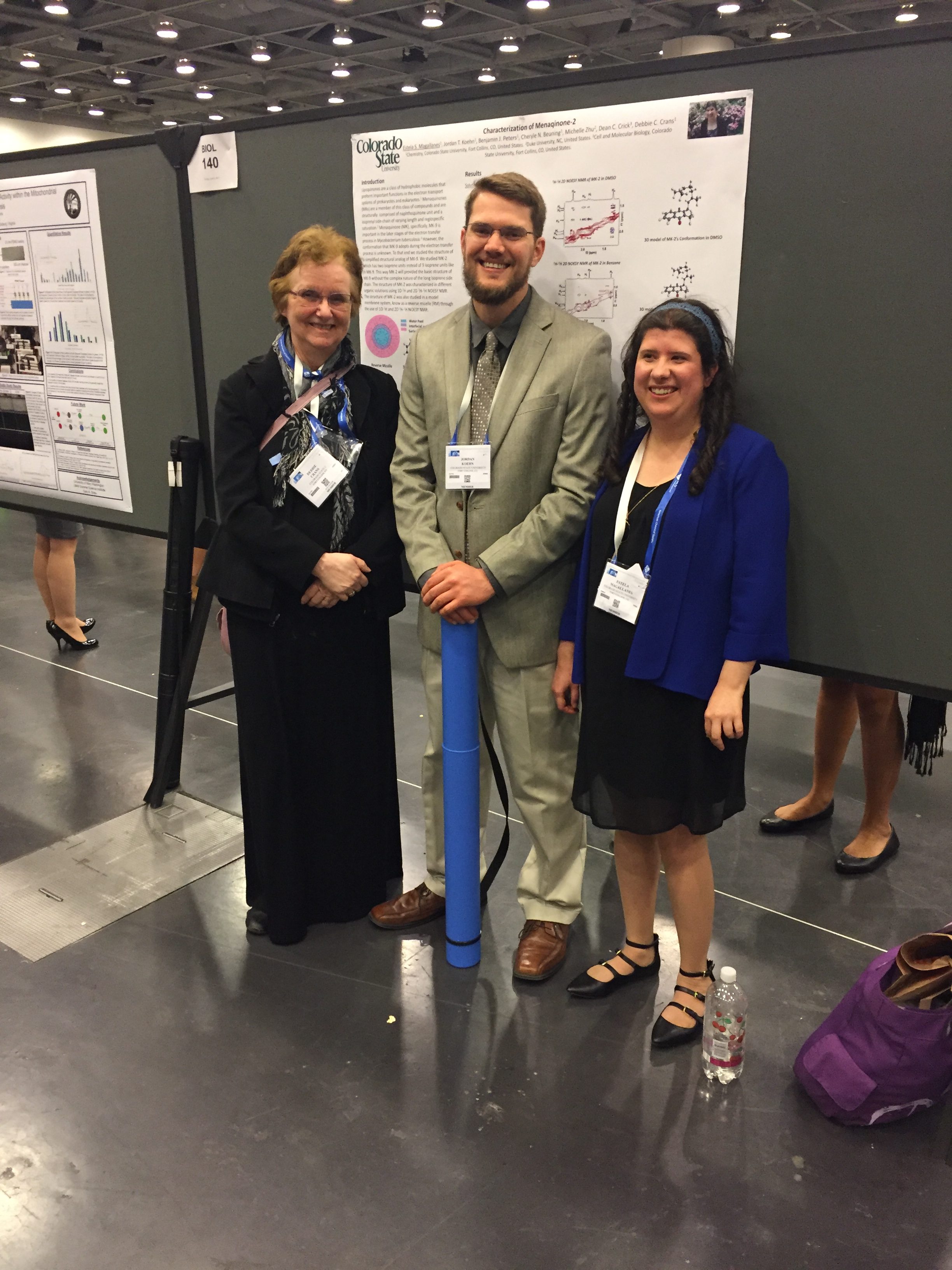
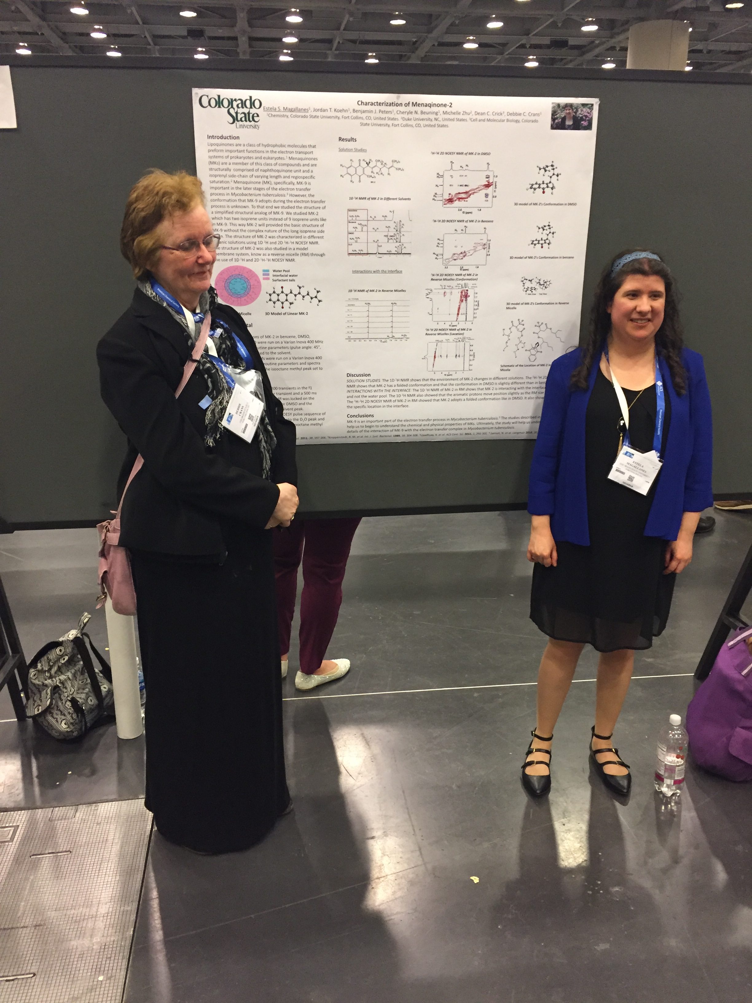
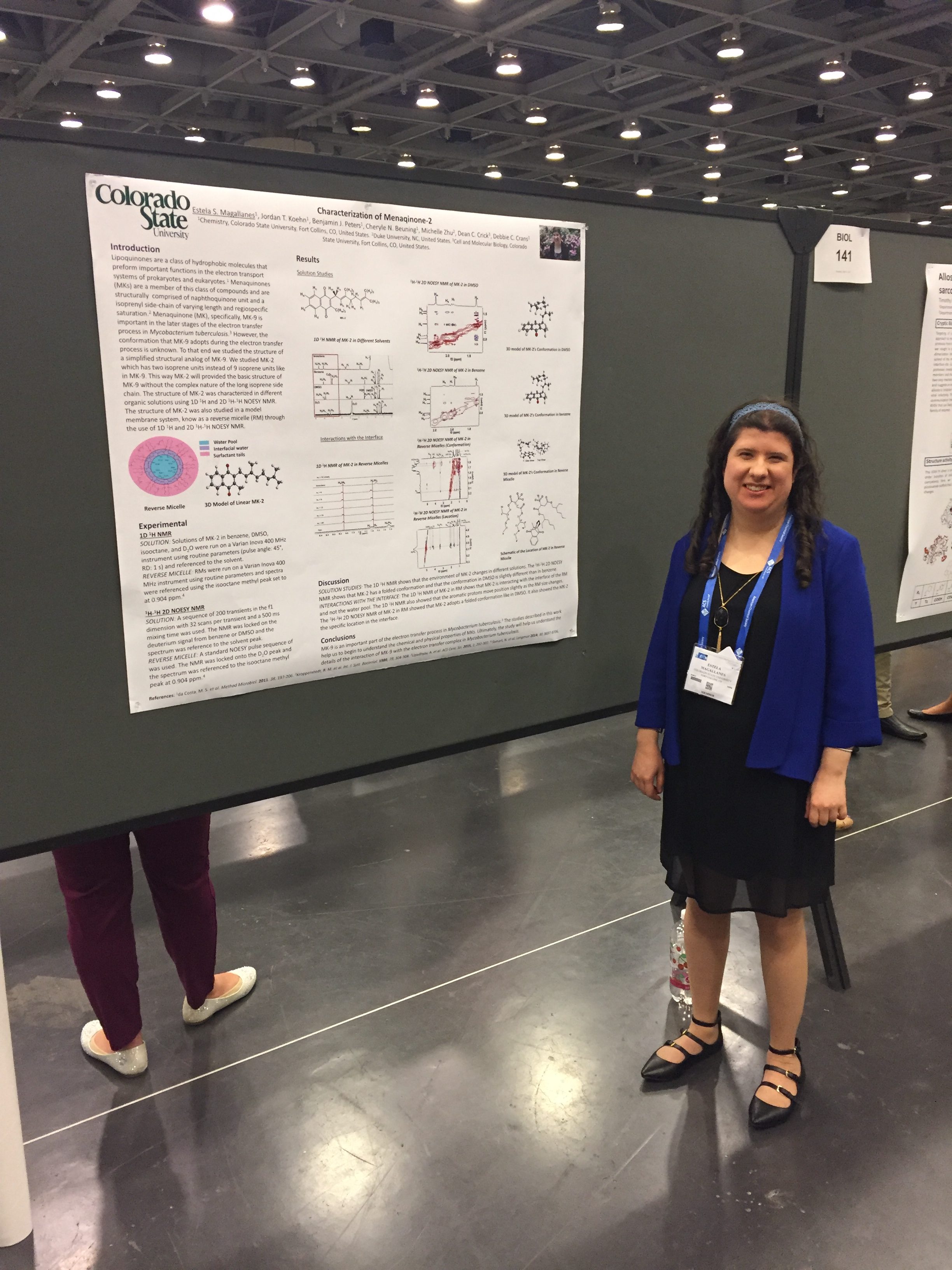
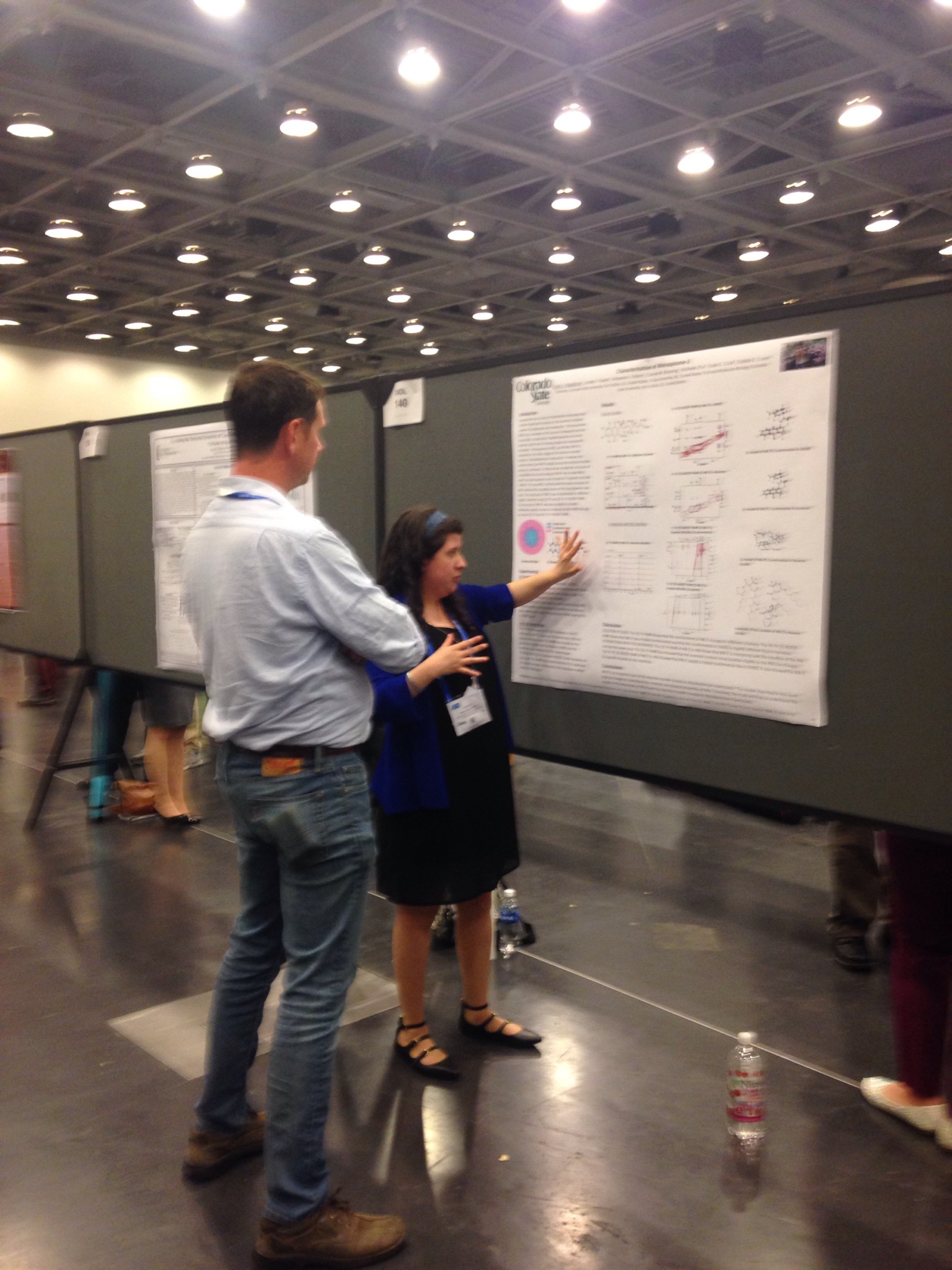
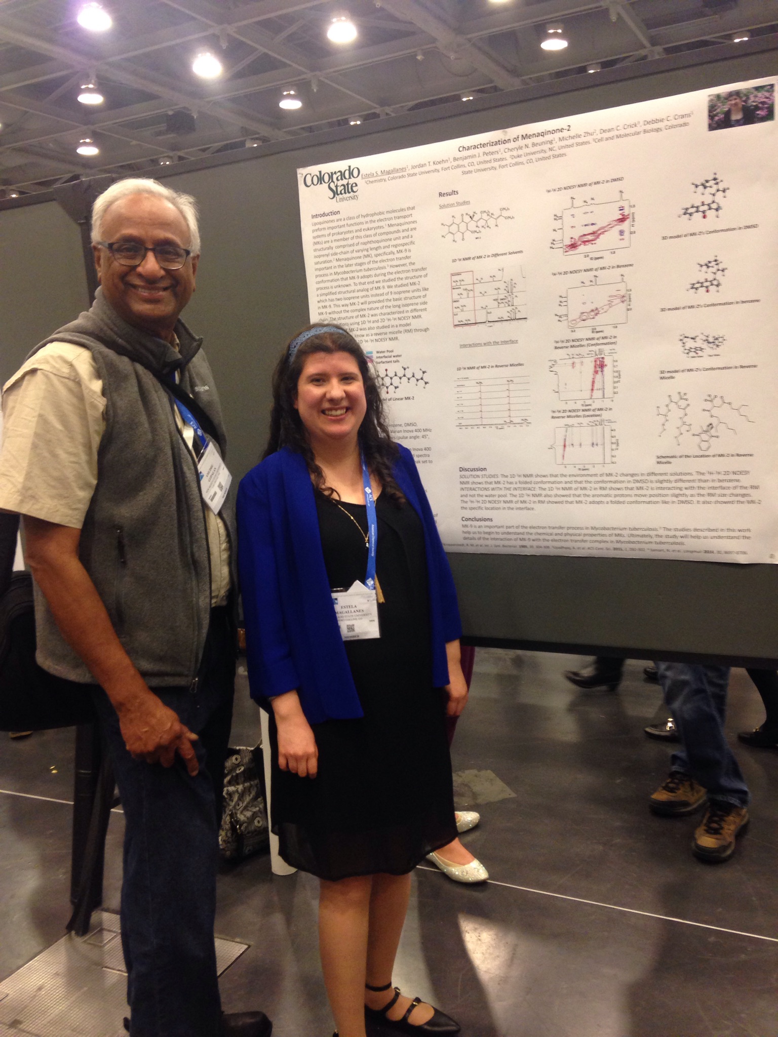
Poster Number and Title: 140 – Characterization of menaqinone-2
Authors: Estela Magallanes, Jordan Koehn, Benjamin Peters, Cheryle Beuning, Michelle Zhu, Dean Crick, Debbie Crans
Authors: Lipoquinones play key roles in the respiratory electron transport systems (ETS) of prokaryotes and eukaryotes contributing to the separation of charge across the membrane by shuttling electrons between protein complexes, acting as electron donors and acceptors. Each of these molecules contains an isoprenyl side-chain of varying length and degree of regiospecific saturation (hydrogenation of double bonds). We have initiated a study of menaquinone-2 (MK-2) with the napthone unit attached to two isoprene units. Specifically, we examined the structure of MK-2 in different organic solvents, and the 1D and 2D 1H NMR spectra suggested that the compound was changing conformation. We also examined the electrochemistry of MK-2 in different solvents. This compound is very insoluble in water we investigated its interaction in a reverse micelle. In these studies, we found that the MK-2 associate with the interface and that the conformation change reflect the favorable interaction with the interface.